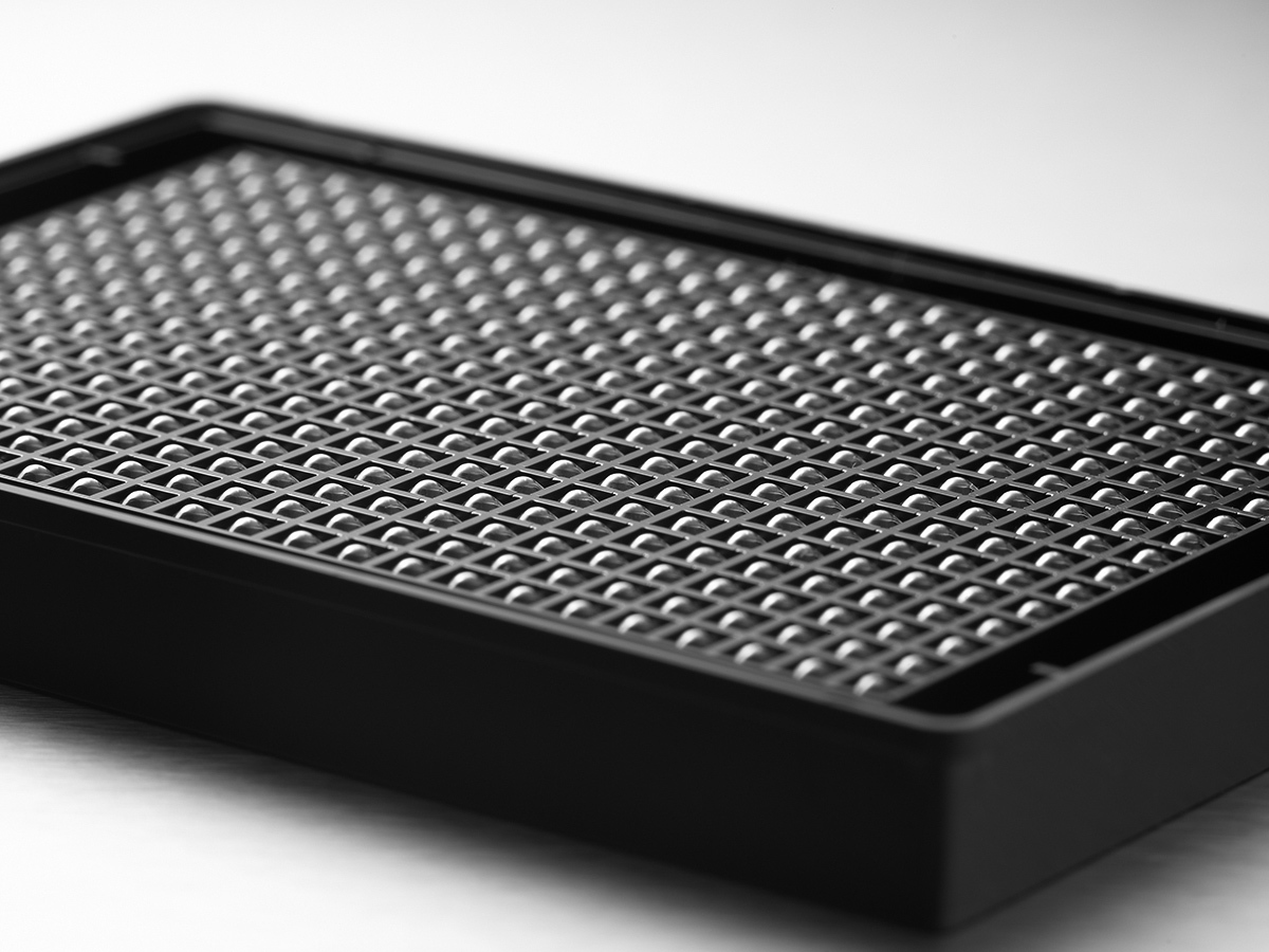
Product Number | 3830 |
Qty./Pk | 10 / Pk |
Qty./Cs | 50 / Cs |
Brand | Corning® |
Plate Format | 384-well |
Plate Feature | Standard |
Plate Color | Clear bottom black |
Well Bottom | Round |
Well Bottom Color | Clear |
Well Volume | 90 µL |
Cell Growth Area | 0.06 cm² (approx.) |
Recommended Medium Well Volume | 20 - 90 µL |
Recommended Working Volume | 20-80 µL |
Surface Treatment | Ultra-Low Attachment |
Sterile | Yes |
Lids Included | Yes |
In this study, Corning spheroid microplates were used for neurosphere formation, proliferation, and migration of hNSCs in an easy-to-use format that is amenable to high throughput screening. The spheroid microplates were used for neurosphere culture of hNSCs over the course of 96 hours, throughout which multipotency was maintained as assessed through Nestin and SOX2 marker expression, followed by subsequent harvesting of neurospheres for differentiation into neuronal, astrocytic, and oligodendrocytic lineages. Imaging of two-dimensional (2D) and 3D cultured hNSCs, in addition to analysis of spheroid size and quantification of migration, was accomplished with the Cytation™ 5 Cell Imaging Multi-Mode Reader and Gen5™ Data Analysis Software (BioTek Instruments, Inc.).
- Application Notes
- American English
- PDF (576.0 kB)
- CLS-AN-334
In this study, A549 cells, a human lung carcinoma cell line, were cultured in spheroid microplates with and without co-culture conditions, including primary human lung fibroblasts [FB] and peripheral blood mononuclear cells [PBMC]. Tumor spheroid viability was screened after treatment with various chemotherapeutics in both single culture and co-culture conditions. Dose-dependent responses of selected chemotherapeutics were compared, demonstrating the impact that including multiple cell types in 3D assays can have on therapeutic outcome.
- Application Notes
- American English
- PDF (732.9 kB)
- CLS-AN-390
Here we demonstrate the use of a 3D neurosphere model, composed of hNSCs, to conduct toxicity testing of potential neurotoxicants. A spheroid microplate was used to create and maintain cells in the 3D model.
- Application Notes
- American English
- PDF (668.1 kB)
- CLS-AN-407
In this study, we combine two novel innovations: Corning HepatoCells and Corning spheroid microplates to create a high throughput 3D model suitable for cytotoxicity screening. Corning HepatoCells are a renewable hepatic cell source derived from primary human hepatocytes. They have consistent phenotypes for cytochrome P450 enzymatic and drug transporter activities, which are necessary for ADME applications such as CYP induction, metabolism, and cytotoxicity studies.
- Application Notes
- American English
- PDF (512.5 kB)
- CLS-DL-AN-340
Here we demonstrate how Corning HepatoCells, an immortalized alternative to primary human hepatocytes can be utilized for a 3D drug screen to discover potential hepatotoxins in conjunction with Corning Spheroid Microplates. These results demonstrate that Corning HepatoCells, together with Corning spheroid microplates, are powerful tools that can be used for reliable and reproducible 3D hepatotoxicity screening.
- Application Notes
- American English
- PDF (458.5 kB)
- CLS-DL-AN-380
- Application Notes
- American English
- PDF (465.4 kB)
- CLS-AN-240
There has been a growing interest in three dimensional (3D) cell culture with more data showing drastic differences in cell behavior and response to drugs when cells are cultured in 3D compared to more traditional two dimensional (2D) models. With the added benefits of 3D systems there come additional challenges in terms of optimization, handling, and assaying. Here we discuss these obstacles and provide recommendations for dealing with them.
- Application Notes
- American English
- PDF (381.6 kB)
- CLS-AN-446
- Application Notes
- American English
- PDF (438.8 kB)
- CLS-AN-447
Formation of gastrointestinal organoids derived from human induced pluripotent stem cells (hiPSC) in Corning® 96-well spheroid microplates in combination with Corning Matrigel® matrix, resulting in a single intestinal organoid in each well.
- Application Notes
- American English
- PDF (1.2 MB)
- CLS-AN-464 A4
Formation of gastrointestinal organoids derived from human induced pluripotent stem cells (hiPSC) in Corning® 96-well spheroid microplates in combination with Corning Matrigel® matrix, resulting in a single intestinal organoid in each well.
- Application Notes
- American English
- PDF (1.2 MB)
- CLS-AN-464
The use of three-dimensional (3D) cell cultures for in vitro drug discovery assays has increased dramatically in recent years because 3D cell culture models more accurately mimic the in vivo environment compared to traditional two-dimensional (2D) monolayer cultures.
- Application Notes
- American English
- PDF (440.7 kB)
- CLS-AN-509
This document summarizes downstream applications and their corresponding techniques as well as equipment for multicellular spheroids generated by Corning spheroid microplate, including monitoring spheroid morphology, viability, proliferation and sort of gene and protein expression etc.
- Application Notes
- American English
- PDF (78.4 kB)
- CLS-AN-545 A4
This document summarizes downstream applications and their corresponding techniques as well as equipment for multicellular spheroids generated by Corning spheroid microplate, including monitoring spheroid morphology, viability, proliferation and sort of gene and protein expression etc.
- Application Notes
- American English
- PDF (81.1 kB)
- CLS-AN-545
Pancreatic ductal adenocarcinoma (PDAC) is a notoriously aggressive tumor type that is typically hypoxic due to oxygen diffusion limitations. Hypoxia can lead to the transcriptional induction of a series of genes, such as GLUT-1 and CA IX, that participate in angiogenesis and tumor growth through hypoxia-inducible factor 1 (HIF-1). To model PDAC in vitro, three-dimensional (3D) cell culture models can be used as they have been shown to develop gradients of diffusion for oxygen and can develop hypoxic cores. In this study, the PDAC cell line PANC-1 was seeded at densities ranging from 500 to 5,000 cells in Corning® spheroid microplates and were cultured for 7 days to form spheroids of varying sizes. RNA was isolated from spheroids at each seeding density, as well as from PANC-1 cells cultured in 2D, using the Promega Maxwell® Rapid Sample Concentrator (RSC) instrument with the Maxwell RSC miRNA Tissue Kit. Relative expression of hypoxia markers HIF-1α, GLUT-1, and CA IX were analyzed relative to 2D culture using RT-qPCR to assess the correlation between spheroid size and hypoxia marker expression. As spheroid density increased, the gene expression of HIF-1α did not change; however, the expression level of HIF-1 targeted genes GLUT-1 and CA IX increased in direct relation to spheroid density.
- Application Notes
- American English
- PDF (296.8 kB)
- CLS-AN-597 A4
Pancreatic ductal adenocarcinoma (PDAC) is a notoriously aggressive tumor type that is typically hypoxic due to oxygen diffusion limitations. Hypoxia can lead to the transcriptional induction of a series of genes, such as GLUT-1 and CA IX, that participate in angiogenesis and tumor growth through hypoxia-inducible factor 1 (HIF-1). To model PDAC in vitro, three-dimensional (3D) cell culture models can be used as they have been shown to develop gradients of diffusion for oxygen and can develop hypoxic cores. In this study, the PDAC cell line PANC-1 was seeded at densities ranging from 500 to 5,000 cells in Corning® spheroid microplates and were cultured for 7 days to form spheroids of varying sizes. RNA was isolated from spheroids at each seeding density, as well as from PANC-1 cells cultured in 2D, using the Promega Maxwell® Rapid Sample Concentrator (RSC) instrument with the Maxwell RSC miRNA Tissue Kit. Relative expression of hypoxia markers HIF-1α, GLUT-1, and CA IX were analyzed relative to 2D culture using RT-qPCR to assess the correlation between spheroid size and hypoxia marker expression. As spheroid density increased, the gene expression of HIF-1α did not change; however, the expression level of HIF-1 targeted genes GLUT-1 and CA IX increased in direct relation to spheroid density.
- Application Notes
- American English
- PDF (309.4 kB)
- CLS-AN-597
- Application Notes
- American English
- PDF (588.5 kB)
- CLS-AN-124
This Customer Application note describes the use of Corning® Ultra-Low Attachment Surface product in the growth and maintenance of spheroid cultures generated from ovarian cancer cells for the investigation of metastasis. The covalently coated hydrogel surface is hydrophilic and uncharged and provides a consistent surface that discourages cell attachment and facilitates the growth of spheroid cultures.
- Application Notes
- American English
- PDF (250.0 kB)
- CLS-AN-127
- Application Notes
- American English
- PDF (244.2 kB)
- CLS-AN-206
Here we compare human liver microtissues (hLiMTs) formed using the hanging drop technique or Corning spheroid microplates and assess the two approaches for their ease of imaging using high content screening.
- Application Notes
- American English
- PDF (373.9 kB)
- CLS-AN-325
Evidence over the past two decades has shown that in vitro three-dimensional (3D) cell models provide more biologically-relevant results than 2D monolayer models for cancer research. This is because cells grown in 3D cultures – like spheroids – more closely resemble the complex microenvironment of a tissue or solid tumor in the human body than 2D cultures. By maintaining physiological traits, such as the natural shape of the cells, inter-cellular communications, cell-matrix interactions, cell proliferation and differentiation, and drug metabolism, 3D models have proven to be useful tools for the screening of anti-cancer drugs.
- Application Notes
- American English
- PDF (2.2 MB)
- Tecan-401631-Spheroids-Fluent-MCA96-V1
Spheroids are organized and functional 3-Dimensional cell aggregates that better mimic the structureof cells in tissues compared to 2D cell culture. This characteristic makes spheroids a powerful tool tostudy more physiologically relevant cell behavior, resulting in more biologically relevant results.
- Citations & References
- American English
- PDF (242.5 kB)
- CLS-AN-507
- Citations & References
- American English
- PDF (1.1 MB)
- Citations & References
- American English
- PDF (1.1 MB)
- Citations & References
- American English
- PDF (529.2 kB)
- Citations & References
- American English
- PDF (1.1 MB)
- Citations & References
- American English
- PDF (1.2 MB)
- CLS-AN-191
- Citations & References
- American English
- PDF (527.4 kB)
- Citations & References
- American English
- PDF (480.5 kB)
- CLS-AN-080
Use the Corning® selection guide to help you choose the best surface for your application and cell type.
- Brochures
- American English
- PDF (774.9 kB)
- CLS-CC-061 REV1
One-page summary of plate and well dimensions for Corning 96, 384, and 1536-well polypropylene and polystyrene microplates. This dimensional information can be used to set up Corning microplates into automation equipment software.
- Drawings
- American English
- PDF (142.8 kB)
- Microplate Dimensions 96, 384, 1536
- FAQS
- American English
- PDF (81.9 kB)
- CLS-DIV-039 EU
- FAQS
- American English
- PDF (80.8 kB)
- CLS-DIV-039
- FAQS
- American English
- PDF (471.7 kB)
- MISC-3D Cell Culutre Flyers-3-15
Updated August 22, 2023
- SDS
- American English
- PDF (50.6 kB)
In this study, a tumor cell line was cultured in the Corning spheroid microplates with and without co-culture conditions, including primary fibroblasts and peripheral blood mononuclear cells. Tumor spheroid viability was screened after treatment with various chemotherapeutics in both single tumor cell line culture and co-culture conditions.
- Posters
- American English
- PDF (5.2 MB)
- CLS-GT-PST-030
Here we demonstrate how Corning HepatoCells, an immortalized alternative to primary human hepatocytes, in conjunction with Corning Spheroid Microplates can be a utilized for a 3D drug screen to discover potential hepatotoxins.
- Posters
- American English
- PDF (2.2 MB)
- CLS-GT-PST-028
In this poster, we demonstrate the use of the spheroid microplate to generate a 3D neurosphere model, composed of hNSCs, to conduct toxicity testing of potential neurotoxicants.
- Posters
- American English
- PDF (1.2 MB)
- Product Information Sheets
- American English
- PDF (642.4 kB)
- CI-Sperhoid Strainer Tip-15
The improved in vivo relevancy of 3D cell culture has driven the adoption of these models in drug discovery. However, the imaging techniques used to characterize these models are highly limited. Due to the thickness and opacity of these 3D cellular structures, current imaging technologies cannot penetrate to the center of the tissues, resulting in only the outer 2 to 3 layers of cells being detected. This causes the dark centers often seen in images of 3D cell culture models, which proves highly problematic for accurate analysis as these outer cells are most exposed to compounds, nutrients and oxygen, and thus do not reflect the entire cell population.
- Product Information Sheets
- American English
- PDF (337.7 kB)
- CLS-AN-024 A4
The improved in vivo relevancy of 3D cell culture has driven the adoption of these models in drug discovery. However, the imaging techniques used to characterize these models are highly limited. Due to the thickness and opacity of these 3D cellular structures, current imaging technologies cannot penetrate to the center of the tissues, resulting in only the outer 2 to 3 layers of cells being detected. This causes the dark centers often seen in images of 3D cell culture models, which proves highly problematic for accurate analysis as these outer cells are most exposed to compounds, nutrients and oxygen, and thus do not reflect the entire cell population.
- Product Information Sheets
- American English
- PDF (339.6 kB)
- CLS-AC-024 REV1
The same Corning Ultra-Low Attachment (ULA) surface that you have used on microplates is now available on a variety of Corning cultureware vessels.
- Product Information Sheets
- American English
- PDF (309.4 kB)
- CLS-CC-013 REV11
Generate, culture, assay, and analyze your spheroids all in one microplate. The Corning spheroid microplate features a novel and proprietary design that allows you to use one microplate to achieve all of these steps without manipulating or transferring delicate spheroids.
- Product Information Sheets
- American English
- PDF (220.2 kB)
- CLS-MP-066 REV2 A4
Generate, culture, assay, and analyze your spheroids all in one microplate. The Corning spheroid microplate features a novel and proprietary design that allows you to use one microplate to achieve all of these steps without manipulating or transferring delicate spheroids.
- Product Information Sheets
- American English
- PDF (211.4 kB)
- CLS-MP-066 REV4
- Product Information Sheets
- American English
- PDF (964.0 kB)
- MISC-3D Cell Culutre Flyers-4-15
In vitro 3D cell culture models are widely recognized as more physiologically relevant systems compared to 2D formats. The 3D models reflect more accurately the complex in vivo microenvironment and have been used in many research areas, such as cancer biology, hepatotoxicity, neurology, pancreatic studies, nephrology, and stem cell biology6.
- Protocols
- American English
- PDF (706.9 kB)
- CLS-AN-235 REV8 A4
In vitro 3D cell culture models are widely recognized as more physiologically relevant systems compared to 2D formats. The 3D models reflect more accurately the complex in vivo microenvironment and have been used in many research areas, such as cancer biology, hepatotoxicity, neurology, pancreatic studies, nephrology, and stem cell biology.
- Protocols
- American English
- PDF (706.7 kB)
- CLS-AN-235 REV8
In vitro 3D cell culture models are widely recognized as more physiologically relevant systems compared to 2D formats. To recapitulate features of native tumor microenvironments, cancer cells can be cultured in Corning spheroid microplates, which combine the Corning Ultra-Low Attachment surface with innovative well geometry to provide an ideal tool for generating, culturing, and assaying 3D multicellular spheroids in the same plate, without the need for a transfer step.
- Protocols
- American English
- PDF (496.9 kB)
- CLS-AN-308 A4
In vitro 3D cell culture models are widely recognized as more physiologically relevant systems compared to 2D formats. To recapitulate features of native tumor microenvironments, cancer cells can be cultured in Corning spheroid microplates, which combine the Corning Ultra-Low Attachment surface with innovative well geometry to provide an ideal tool for generating, culturing, and assaying 3D multicellular spheroids in the same plate, without the need for a transfer step.
- Protocols
- American English
- PDF (496.8 kB)
- CLS-AN-308 REV2
- Product Descriptions
- American English
- Quality Certificate - Lot No.35524032
- Quality Certificate - Lot No.32624029
- Quality Certificate - Lot No.32424016
- Quality Certificate - Lot No.28524039
- Quality Certificate - Lot No.27124008
- Quality Certificate - Lot No.26424008
- Quality Certificate - Lot No.25724004
- Quality Certificate - Lot No.24324022
- Quality Certificate - Lot No.23624022
- Quality Certificate - Lot No.23524019
- Quality Certificate - Lot No.22924024
- Quality Certificate - Lot No.08824045
- Quality Certificate - Lot No.06024044
- Quality Certificate - Lot No.05924011
- Quality Certificate - Lot No.02624002
- Quality Certificate - Lot No.02424052
- Quality Certificate - Lot No.01024004
- Quality Certificate - Lot No.31823042
- Quality Certificate - Lot No.29723010
- Quality Certificate - Lot No.26523022
- Quality Certificate - Lot No.25623044
- Quality Certificate - Lot No.24123031
- Quality Certificate - Lot No.06823011